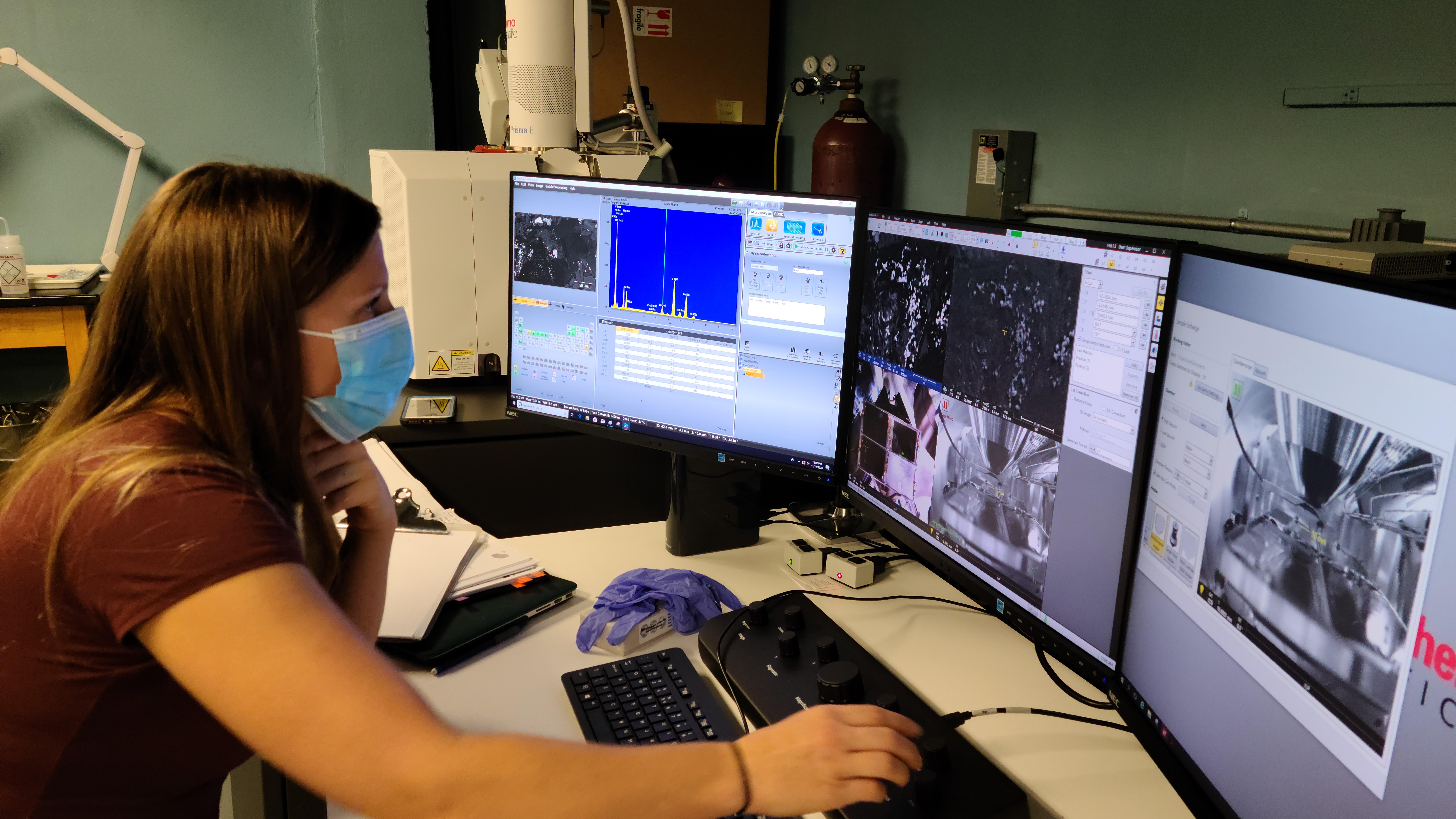
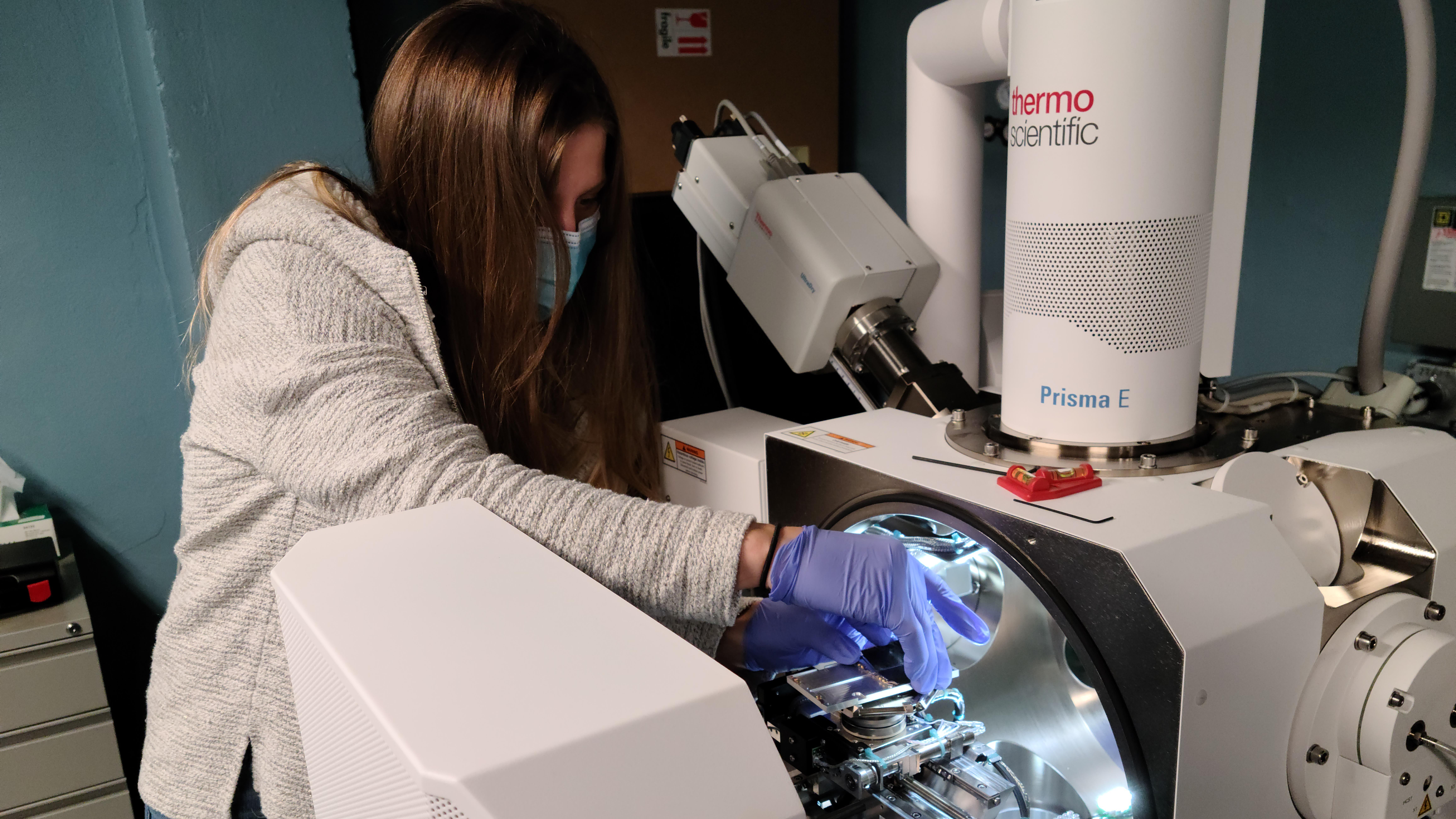
Student Jennifer Miller uses a scanning electron microscope to analyze rock samples for more accurate dating.
How old do you think that rock is?
Geology major Jennifer Miller is working on an answer.
As part of her senior research project, Miller traveled to Indiana University of Pennsylvania with samples she collected from a vertical igneous rock layer in Adah, Pa.
There, she used the school’s scanning electron microscope to analyze the composition, looking for the presence of apatite, a calcium phosphate mineral.
The rock was likely emplaced when Pangaea, a supercontinent that existed in the late Paleozoic and early Mesozoic eras, broke apart. But providing a specific age of the rock has been a challenge.
“Jen verified the presence of apatite in the rock using the SEM,” explained Dr. Daniel Harris, geology professor and Miller’s research supervisor. “We can then extract that mineral and use it to provide a better crystallization age, which then would provide a better estimate of the relationship of the rock to Pangaea breakup.”
The next step will be to extract the apatite and visit a geochronology lab for further examination.
“She got some excellent data,” Harris said. “This will help us to better understand this intrusion.”


